THE LIMITATIONS OF DIAGNOSING DRUG-RESISTANT EPILEPSY
Surgery is the only curative treatment for a third of patients with
drug-resistant focal epilepsy.
Precise prior localisation of the epileptogenic zone is crucial to the success of surgery, but it is a
delicate operation. To achieve this, doctors currently use a combination of information:
-
video recordings coupled with
electroencephalograms ;
-
moprhological data obtained by magnetic resonance imaging (MRI) ;
- information on
metabolically active areas of the brain, obtained using [18F]FDG positron emission tomography
(PET)
In 60% of cases, this pre-surgical assessment phase makes it possible to identify epileptogenic areas with a high degree of confidence and propose immediate surgery. The rest of the time, this is not possible, particularly when [18F]FDG PET is not sufficiently informative.
DPA-714, an additional tool
The [18F]DPA-714 radiotracer has recently been proposed as an interesting alternative to FDG. DPA-714 is a ligand for the TSPO protein, which is overproduced by activated glial cells and astrocytes during neuroinflammation, a phenomenon that occurs notably in epilepsy.
In a trial involving 23 people, the BioMaps laboratory (SHFJ), a leader in the use of [18F]DPA-714 in preclinical and clinical studies, has shown that [18F]DPA-714 PET provides more relevant information on the location of epileptogenic foci than [18F]FDG PET. This benefit has made it possible to surgically manage patients who would not have benefited from surgery with the results of FDG PET.
These results indicate that [18F]DPA-714 PET could be an additional tool for the best possible localisation of epileptogenic foci, particularly for patients for whom FDG PET is not informative.
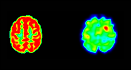
PET scan of a patient with a suspected right pericentral epileptogenic focus. The [18F]DPA-714 PET scan (right) shows a strong and easily perceptible focal increase in uptake in the right precentral zone. Retrospective analysis of [18F]FDG PET (left) reveals sulcal hypometabolism in the region indicated by [18F]DPA-714 PET. (c) Cheval et al., Neurology.
Contact :